Background and Purpose
Quantitative tissue characteristics derived from medical images, also called radiomics, contain valuable prognostic information in several tumour-sites. The large number of features available increases the risk of overfitting. Typically test-retest CT-scans are used to reduce dimensionality and select robust features. However, these scans are not always available. We propose to use different phases of respiratory-correlated 4D CT-scans (4DCT) as alternative.
Materials and Methods
In test-retest CT-scans of 26 non-small cell lung cancer (NSCLC) patients and 4DCT-scans (8 breathing phases) of 20 NSCLC and 20 oesophageal cancer patients, 1045 radiomics features of the primary tumours were calculated. A concordance correlation coefficient (CCC) >0.85 was used to identify robust features. Correlation with prognostic value was tested using univariate cox regression in 120 oesophageal cancer patients.
Results
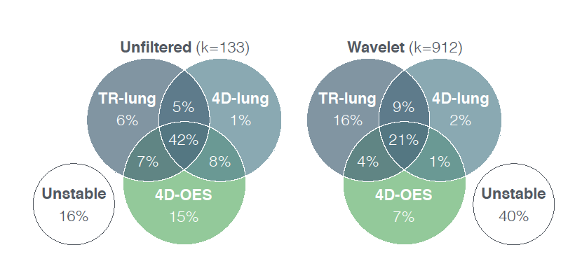
Features based on unfiltered images demonstrated greater robustness than wavelet-filtered features. In total 63/74 (85%) unfiltered features and 268/299 (90%) wavelet features stable in the 4D-lung dataset were also stable in the test-retest dataset. In oesophageal cancer 397/1045 (38%) features were robust, of which 108 features were significantly associated with overall-survival.
Conclusion
4DCT-scans can be used as alternative to eliminate unstable radiomics features as first step in a feature selection procedure. Feature robustness is tumour-site specific and independent of prognostic value.

Venn chart visualizing the overlap of stable features with CCC>0.85 in the test-retest lung (TR-lung), 4D-lung and 4D-oesophagus (4D-OES) dataset respectively.
| 