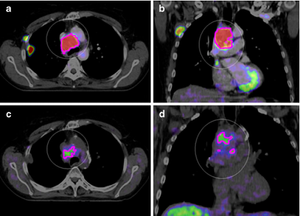
PET imaging of hypoxia using [18F]HX4: a phase I trial

TitlePET imaging of hypoxia using [18F]HX4: a phase I trial
Publication TypeJournal Article
Year of Publication2010
AuthorsLoon, J, Janssen, MHM, Oellers, MC, Aerts, HJWL, Dubois, LJ, Hochstenbag, M, Dingemans, A-MC, Lalisang, R, Brans, B, Windhorst, B, Dongen, GA, Kolb, HC, Zhang, J, De Ruysscher, D, Lambin, P
JournalEuropean Journal of Nuclear Medicine and Molecular Imaging
Volume37
Issue9
Pagination1663 - 1668
Date PublishedJan-08-2010
Publication Languageeng
ISSN1619-7070
Keywords2-Nitroimidazoles, HX4, Hypoxia, PET
Abstract


Download the images using these instructions and this DOI : 10.1007/s00259-010-1437-x


Background and purpose
Non-invasive PET imaging of tumour hypoxia could help in the selection of those patients who could benefit from chemotherapy or radiation with specific antihypoxic treatments such as bioreductive drugs or hypoxic radiosensitizers. In this phase I trial, we aimed to determine the toxicity of [18F]HX4, a member of the 2-nitroimidazole family, at different dose levels. The secondary aim was to analyse image quality related to the HX4 dose and the timing of imaging.

Methods
Patients with a histologically proven solid cancer without curative treatment options were eligible for this study. A study design with two dose steps was used in which a single dose of a maximum of 222MBq (step 1) or 444 MBq (step 2) [18F]HX4 was injected. Toxicity was scored on day 0 and on days 3 and 7 after injection, according to the CTCAE 3.0 scoring system. PET/CT images of the largest tumour site were acquired 30, 60 and 120 min after injection.

Results
Six patients with stage IV carcinoma were included, four with non-small-cell lung carcinoma, one with thymus carcinoma, and one with colon carcinoma. No toxicity was observed in any of the patients at either dose level. The median tumour to muscle ratio 120 min after injection was 1.40 (range 0.63–1.98).

Conclusion 
The findings of this study showed that [18F] HX4 PET imaging for the detection of hypoxia is not associated with any toxicity. Imaging was successful; however, future trials are needed to determine the optimal image parameters.

URLhttp://link.springer.com/10.1007/s00259-010-1437-x
DOI10.1007/s00259-010-1437-x
Short TitleEur J Nucl Med Mol Imaging