· David Hofstede , Dario Di Perri , Erik Roelofs , Alida Postma , Esther Troost , Daniëlle Eekers · Tutorial · 10.17195/candat.2022.02.1
Caudate Nucleus
Contouring instructions for the caudate nucleus.
Caudate Nucleus
The caudate nucleus is a major component of the basal ganglia and is involved in much more than motor control alone. In addition to its role in movement planning and coordination, the caudate nucleus contributes to cognitive processes such as attention, learning, executive functioning, memory, motivation, and goal-directed behaviour. Through its extensive connections with the frontal cortex and limbic system, it also plays a role in emotional processing and reward-related functions.Because of its involvement in cognitive and behavioural networks, there is increasing interest in understanding the effects of radiation dose to the caudate nucleus and its potential contribution to neurocognitive outcomes and quality of life after brain irradiation. Accurate delineation is therefore important for both clinical practice and research.
For contouring, the left and right caudate nucleus should be delineated separately. Co-registration with a T1-weighted MRI is recommended to facilitate accurate identification of the structure. The caudate nucleus is a curved, C-shaped gray matter structure.
Three anatomical components:
- Head – the large anterior portion.
- Body – the elongated central segment.
- Tail – a thin posterior extension that follows the curvature of the lateral ventricle.
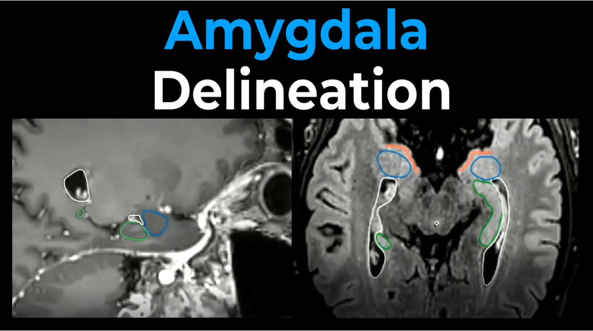
The head of the caudate nucleus forms the lateral wall of the anterior horn of the lateral ventricle. Inferiorly, it is continuous with the nucleus accumbens, while anterolaterally it is closely related to the putamen. The body of the caudate nucleus lies along the lateral aspect of the lateral ventricle and is separated from the putamen by the internal capsule.Posteriorly, the tail curves around the thalamus and follows the roof of the temporal horn of the lateral ventricle, eventually extending towards the amygdala. The most distal part of the tail can be difficult to identify on routine clinical MRI scans and may therefore be less reliably visualized.
The caudate nucleus, together with the putamen, forms the striatum, a central structure within the cortico-striatal networks that support motor, cognitive, and emotional functioning.
The accompanying video provides a step-by-step demonstration of how to identify and contour the caudate nucleus using anatomical landmarks on MRI. This educational resource is intended to support consistent, accurate, and reproducible delineation for clinical and research applications.
Please site as follows:
Hofstede D, Di Perri D, Roelofs E, Postma AA, Troost EGC, Eekers DBP. EPTN International Neurological Contouring Atlas (INCA) instruction videos. CancerData, 2022. doi:10.17195/candat.2022.02.1